Ciencias de la Salud- Caso clínico
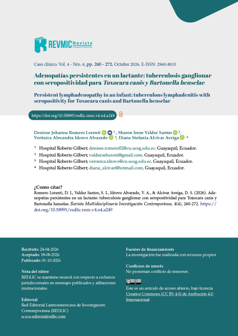
Persistent lymphadenopathy in an infant: tuberculous lymphadenitis with seropositivity for Toxocara canis and Bartonella henselae
Adenopatías persistentes en un lactante: tuberculosis ganglionar con seropositividad para Toxocara canis y Bartonella henselae
Abstract
Persistent lymphadenopathy in pediatrics represents a diagnostic challenge due to the wide range of infectious etiologies involved; extrapulmonary tuberculosis accounts for 20–30% of pediatric tuberculosis cases, with tuberculous lymphadenitis being one of its most common forms. We present the case of a 1-year-4-month-old male infant with persistent fever, pruritic skin lesions, and progressive cervical and inguinal lymphadenopathy unresponsive to antibiotic therapy. Laboratory tests showed leukocytosis, thrombocytosis, and cervical ultrasound findings of nodal necrosis. Lymph node biopsy revealed caseous and purulent material, and tuberculosis lymphadenitis was confirmed by PCR/DNA testing of the lymph node and purulent secretion. Additionally, seropositivity for Toxocara canis and Bartonella henselae was documented, broadening the differential diagnosis. The patient received anti-tuberculosis therapy and albendazole, with favorable clinical evolution. This case highlights the importance of a syndromic approach and early microbiological confirmation in persistent pediatric lymphadenopathy.
Las adenopatías persistentes en pediatría representan un reto diagnóstico debido a la diversidad de etiologías infecciosas involucradas; la tuberculosis extrapulmonar corresponde al 20–30% de los casos pediátricos, siendo la linfadenitis tuberculosa una de sus formas más frecuentes. Se presenta un lactante masculino de 1 año y 4 meses con fiebre persistente, lesiones cutáneas pruriginosas y adenopatías cervicales e inguinales progresivas sin respuesta a antibióticos. Los estudios evidenciaron leucocitosis, trombocitosis y ecografía cervical con necrosis ganglionar. La biopsia mostró material caseoso y purulento, confirmándose tuberculosis ganglionar mediante PCR/ADN en ganglio y secreción. Adicionalmente, se documentó seropositividad para Toxocara canis y Bartonella henselae, ampliando el diagnóstico diferencial. El paciente recibió tratamiento antifímico y albendazol, con evolución favorable. Este caso resalta la importancia del abordaje sindrómico y de la confirmación microbiológica temprana en adenopatías persistentes pediátricas.
Keywords
- persistent lymphadenopathy
- tuberculous lymphadenitis
- toxocariasis
- bartonellosis
- infant
- pediatrics
References
Deosthali A, Donches K, DelVecchio M, Aronoff S. Etiologies of Pediatric Cervical Lymphadenopathy: A Systematic Review of 2687 Subjects. Global Pediatric Health. 2019;6:2333794X19865440. DOI: https://doi.org/10.1177/2333794X19865440
World Health Organization. Global tuberculosis report 2024. Geneva: World Health Organization; 2024. Disponible en: https://www.who.int/teams/global-tuberculosis-programme/tb-reports/global-tuberculosis-report-2024?utm_source=chatgpt.com
Ministerio de Salud Pública del Ecuador. Tratamiento de la infección por tuberculosis, tuberculosis sensible y resistente: Guía de Práctica Clínica. Quito: MSP; 2024. Disponible en: Guía de Práctica Clínica Tuberculosis Ecuador 2024
Yu TZ, Pang Y, Du J, et al. Diagnostic value of ultrasound in cervical tuberculous lymphadenitis. Quantitative Imaging in Medicine and Surgery. 2021;11(6):2587-2598. DOI: https://doi.org/10.21037/qims-20-842
Öcal Demir S, Bayhan GI, Tanır G. Cat scratch disease and differential diagnosis in persistent lymphadenopathy in children. Children. 2024;11(3):310. DOI: https://doi.org/10.3390/children11030310
Rostami A, Riahi SM, Holland CV, et al. Seroprevalence estimates for toxocariasis in people worldwide: A systematic review and meta-analysis. PLoS Neglected Tropical Diseases. 2019;13(12):e0007809. DOI: https://doi.org/10.1371/journal.pntd.0007809
Chen J, Zhou DH, Nisbet AJ, et al. Advances in molecular identification, taxonomy, genetic variation and diagnosis of Toxocara spp. Infectious Diseases of Poverty. 2020;9(1):85. DOI: https://doi.org/10.1186/s40249-020-00692-2
Florin TA, Zaoutis TE, Zaoutis LB. Beyond cat scratch disease: widening spectrum of Bartonella henselae infection. Pediatrics. 2020;146(4):e20200018. DOI: https://doi.org/10.1542/peds.2020-0018
Kay AW, Verkuijl SE, et al. Xpert MTB/RIF Ultra assay for tuberculosis disease and rifampicin resistance in children. Cochrane Database of Systematic Reviews. 2025;1(1):CD013359. DOI: https://doi.org/10.1002/14651858.CD013359.pub3
Harris JE, Patel NN, Wai K, Rosbe KW. Management of Pediatric Persistent Asymptomatic Cervical Lymphadenopathy. Otolaryngology–Head and Neck Surgery. 2024;170(1):69-75. DOI: https://doi.org/10.1002/ohn.420
Pecora F, Abate L, Scavone S, Petrucci I, Costa F, Caminiti C, et al. Management of Infectious Lymphadenitis in Children. Children. 2021;8(10):860. DOI: https://doi.org/10.3390/children8100860
Baykan AH, Sayiner HS, Aydin E, Koc M, Inan I, Erturk SM. Extrapulmonary tuberculosis: an old but resurgent problem. Insights into Imaging. 2022;13(1):39. DOI: https://doi.org/10.1186/s13244-022-01172-0
Kumar N, Khan A, Boora S, et al. Diagnosis of tuberculous lymphadenitis by molecular and immunological tools. Medical Microecology. 2024;22:100116. DOI: https://doi.org/10.1016/j.medmic.2024.100116
Martinez L, Cords O, Horsburgh CR Jr, Andrews JR; Pediatric TB Contact Studies Consortium. The risk of tuberculosis in children after close exposure: a systematic review and individual-participant meta-analysis. Lancet. 2020;395(10228):973-984. DOI: https://doi.org/10.1016/S0140-6736(20)30166-5
Magnaval JF, Bouhsira E, Fillaux J. Therapy and Prevention for Human Toxocariasis. Microorganisms. 2022;10(2):241. DOI: https://doi.org/10.3390/microorganisms10020241
Deregibus MI, Bagnara EI, Buchovsky A. Cat-scratch disease: experience in a tertiary care children’s hospital. Archivos Argentinos de Pediatria. 2023;121(1):e202202592. DOI: https://doi.org/10.5546/aap.2022-02592
Nawrocki CC, Max RJ, Marzec NS, Nelson CA. Atypical Manifestations of Cat-Scratch Disease, United States, 2005-2014. Emerging Infectious Diseases. 2020;26(7):1438-1446. DOI: https://doi.org/10.3201/eid2607.200034
Fusani L, Venturini E, Chiappini E, Galli L. Cat scratch disease in childhood: a 10-year experience of an Italian tertiary care children’s hospital. World Journal of Pediatrics. 2023;19(2):189-193. DOI: https://doi.org/10.1007/s12519-022-00616-3
Hermans SM, et al. Post-tuberculosis treatment paradoxical reactions. Infection. 2024;52(5):2083-2095. DOI: https://doi.org/10.1007/s15010-024-02310-0
Cunningham ET Jr, Zierhut M. Ocular Toxocariasis. Ocular Immunology and Inflammation. 2021;29(7-8):1243-1245. DOI: https://doi.org/10.1080/09273948.2021.2018825
Published
History
- Received
- 12 Jul 2026
- Accepted
- 16 Aug 2026
- Published
- 1 Oct 2026
Metrics
110 downloads
last 7 months
Issue
Section
How to Cite
License
Copyright (c) 2026 Denisse Johanna Romero Lorenti, Sharon Irene Valdez Santos, Verónica Alexandra Idrovo Alvarado, Diana Stefanía Alcívar Aveiga. Los autores que publiquen en la revista Multidisciplinaria Investigación Contemporánea aceptan los siguientes términos: 1. Los autores mantienen sus derechos de autor (copyright) y otorgan a la revista Multidisciplinaria Investigación Contemporánea el derecho de la primera publicación de su trabajo, bajo una licencia Creative Commons Attribution 4.0. Esta licencia permite que terceros utilicen el contenido publicado, siempre que se mencione la autoría y la primera publicación en esta revista. 2. Los autores pueden establecer acuerdos adicionales para la distribución no exclusiva de la versión publicada de su artículo en otros lugares, como repositorios institucionales, siempre y cuando se indique claramente que el trabajo fue publicado por primera vez en esta revista. 3. Los autores conservan sus derechos de autor (copyright) y garantizan a la revista Multidisciplinaria Investigación Contemporánea el derecho a publicar el manuscrito a través de los canales que considere apropiados. 4. Se permite y se recomienda a los autores compartir su trabajo en línea (por ejemplo, en repositorios institucionales o páginas web personales), una vez que el manuscrito haya sido aceptado para su publicación. Esto puede conducir a intercambios productivos y a una mayor y más rápida citación del trabajo publicado. Detalle de Licencia CC BY (https://creativecommons.org/licenses/by/4.0/) Usted es libre de: Compartir — copiar y redistribuir el material en cualquier medio o formato para cualquier propósito, incluso comercialmente. Adaptar — remezclar, transformar y construir a partir del material para cualquier propósito, incluso comercialmente. La licenciante no puede revocar estas libertades en tanto usted siga los términos de la licencia Bajo los siguientes términos: Atribución — Usted debe dar crédito de manera adecuada , brindar un enlace a la licencia, e indicar si se han realizado cambios . Puede hacerlo en cualquier forma razonable, pero no de forma tal que sugiera que usted o su uso tienen el apoyo de la licenciante. No hay restricciones adicionales — No puede aplicar términos legales ni medidas tecnológicas que restrinjan legalmente a otras a hacer cualquier uso permitido por la licencia.

This work is licensed under a Creative Commons Attribution 4.0 International License.
Los autores que publiquen en la revista Multidisciplinaria Investigación Contemporánea aceptan los siguientes términos:
- Los autores mantienen sus derechos de autor (copyright) y otorgan a la revista Multidisciplinaria Investigación Contemporánea el derecho de la primera publicación de su trabajo, bajo una licencia Creative Commons Attribution 4.0. Esta licencia permite que terceros utilicen el contenido publicado, siempre que se mencione la autoría y la primera publicación en esta revista.
- Los autores pueden establecer acuerdos adicionales para la distribución no exclusiva de la versión publicada de su artículo en otros lugares, como repositorios institucionales, siempre y cuando se indique claramente que el trabajo fue publicado por primera vez en esta revista.
- Los autores conservan sus derechos de autor (copyright) y garantizan a la revista Multidisciplinaria Investigación Contemporánea el derecho a publicar el manuscrito a través de los canales que considere apropiados.
- Se permite y se recomienda a los autores compartir su trabajo en línea (por ejemplo, en repositorios institucionales o páginas web personales), una vez que el manuscrito haya sido aceptado para su publicación. Esto puede conducir a intercambios productivos y a una mayor y más rápida citación del trabajo publicado.
Detalle de Licencia CC BY (https://creativecommons.org/licenses/by/4.0/)
Usted es libre de:
- Compartir — copiar y redistribuir el material en cualquier medio o formato para cualquier propósito, incluso comercialmente.
- Adaptar — remezclar, transformar y construir a partir del material para cualquier propósito, incluso comercialmente.
- La licenciante no puede revocar estas libertades en tanto usted siga los términos de la licencia
Bajo los siguientes términos:
- Atribución — Usted debe dar crédito de manera adecuada , brindar un enlace a la licencia, e indicar si se han realizado cambios . Puede hacerlo en cualquier forma razonable, pero no de forma tal que sugiera que usted o su uso tienen el apoyo de la licenciante.
- No hay restricciones adicionales — No puede aplicar términos legales ni medidas tecnológicas que restrinjan legalmente a otras a hacer cualquier uso permitido por la licencia.